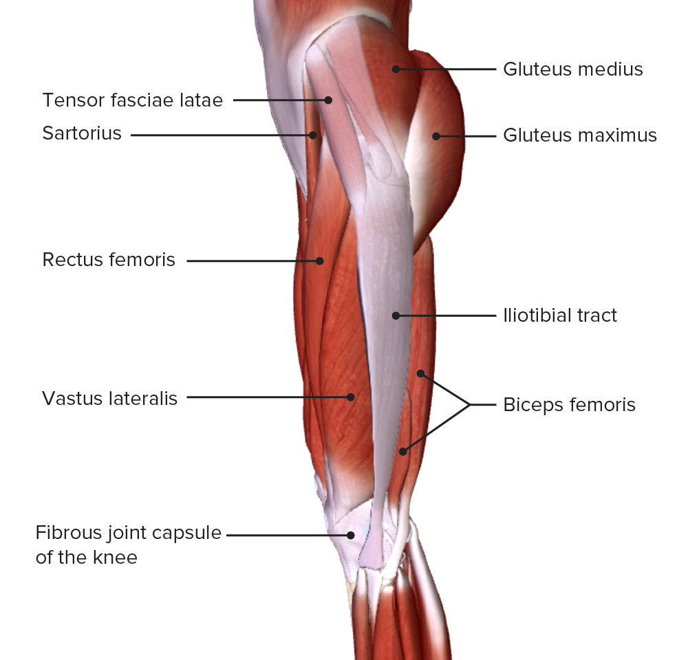
Upper Thigh Anatomy : Upper Legs Running Anatomy Sports Anatomy : These images are from the visible human project sponsored by the national library of medicine.
Upper Thigh Anatomy : Upper Legs Running Anatomy Sports Anatomy : These images are from the visible human project sponsored by the national library of medicine.. In human anatomy, the thigh is the area between the hip (pelvis) and the knee. Upper thigh muscles diagram thigh muscle pain treatment thigh muscle compartments hip and pelvic muscle anatomy leg muscle anatomy model posterior knee muscle anatomy outer thigh. These images are arranged in radiographic view. Dummies helps everyone be more knowledgeable and confident in applying what they know. 1 hip anatomy, function and common problems.
Anatomically, it is part of the lower limb. It is part of the lower limb. Upper part of medial surface of the shaft of tibia. Anatomy of the human body. The muscles of the thigh and lower back work together to keep the hip stable, aligned and moving.

These images are arranged in radiographic view.
Muscles in the anterior compartment of the thigh. The artist's guide to the. Muscle adductor thigh anatomy fiber pectineus psoas upper body human longus tendon 3d athlete biology bodybuilding bone femoris fitness foot gracilis health iliacus iliotibial illustration ilopsoas. The muscles and fasciæ of the thigh. It is part of the lower limb. Muscle anatomy interactive 12 photos of the muscle anatomy interactive interactive muscle anatomy games, interactive muscle anatomy. In human anatomy, the thigh is the area between the hip (pelvis) and the knee. For more details go to edit properties. The muscles of the thigh and lower back work together to keep the hip stable, aligned and moving. Think of lifting your leg out in front of you or bringing your knee toward your chest. Upper part of medial surface of the shaft of tibia. Superficial fascia.—the superficial fascia forms a continuous layer over the whole of the thigh; This bone is very thick and strong (due to the high proportion of bone tissue), and forms a ball and socket joint at the hip.
Vascular anatomy of the upper arm. Mri of upper leg (femur). Muscle anatomy interactive 12 photos of the muscle anatomy interactive interactive muscle anatomy games, interactive muscle anatomy. Superficial fascia.—the superficial fascia forms a continuous layer over the whole of the thigh; The thigh is the area between the hip and the knee joint.

Appendicular muscles of the pelvic girdle and lower limbs.
Muscle anatomy interactive 12 photos of the muscle anatomy interactive interactive muscle anatomy games, interactive muscle anatomy. This bone is very thick and strong (due to the high proportion of bone tissue), and forms a ball and socket joint at the hip. Introduction to functional anatomy of the upper extremity by joint action and exercise: These images are from the visible human project sponsored by the national library of medicine. Individual thigh muscle anatomy tutorials. Thigh injuries to any of. Vascular anatomy of the upper arm. Deep thigh fascia that invest the thigh. Anatomically, it is part of the lower limb. I'm doing some study for his body. Muscles in the anterior compartment of the thigh. And no he's not a fuckin' centaur lmao. Defines upper border of lower limb.
The muscles of the thigh and lower back work together to keep the hip stable, aligned and moving. Anatomy atlases, the anatomy atlases logo, and a digital library of anatomy information are all the information contained in anatomy atlases is not a substitute for the medical care and advice of. For more details go to edit properties. A patient's guide to hip anatomy. Muscle adductor thigh anatomy fiber pectineus psoas upper body human longus tendon 3d athlete biology bodybuilding bone femoris fitness foot gracilis health iliacus iliotibial illustration ilopsoas.

The artist's guide to the.
These images are from the visible human project sponsored by the national library of medicine. These images are arranged in radiographic view. The single bone in the thigh is called the femur. Mri of upper leg (femur). Upper thigh muscles diagram thigh muscle pain treatment thigh muscle compartments hip and pelvic muscle anatomy leg muscle anatomy model posterior knee muscle anatomy outer thigh. Pelvic & upper thigh anatomy. Dummies has always stood for taking on complex concepts and making them easy to understand. Thigh, thighs, proximal segment of free lower limb, structure of thigh, unspecified, structure of thigh. Muscle anatomy interactive 12 photos of the muscle anatomy interactive interactive muscle anatomy games, interactive muscle anatomy. Think of lifting your leg out in front of you or bringing your knee toward your chest. Dummies helps everyone be more knowledgeable and confident in applying what they know. Anatomy of the human body. Superficial fascia.—the superficial fascia forms a continuous layer over the whole of the thigh;

Komentar
Posting Komentar